Bone Scan
A bone scan is a nuclear imaging technique of the bone. It can help diagnose a number of bone conditions, including cancer of the bone or metastasis, location of bone inflammation and fractures (that may not be visible in traditional X-ray images), and bone infection (osteomyelitis).
It provides functional imaging and allows visualization of bone metabolism or bone remodeling, which most other imaging techniques (such as X-ray computed tomography, CT) cannot. Bone scan competes with positron emission tomography (PET) for imaging of abnormal metabolism in bones, but is considerably less expensive. Bone scan has higher sensitivity but lower specificity than CT or MRI for diagnosis of scaphoid fractures following negative plain radiography.

A bone scan is a test that uses nuclear imaging to helps diagnose and track several types of bone disease. Nuclear imaging involves using small amounts of radioactive substances (radiotracers), a special camera that can detect the radioactivity and a computer to see structures such as bones inside the body.
A bone scan might help determine the cause of unexplained bone pain. The test is sensitive to differences in bone metabolism, which are highlighted in the body by the radioactive tracer. Scanning the whole skeleton helps in diagnosing a wide range of bone disorders, including:
- Fractures
- Arthritis
- Paget’s disease of bone
- Cancer originating in bone
- Cancer that has metastasized to bone from a different site
- Infection of the joints, joint replacements or bones
Preparation for Bone Scan
You typically don’t need to restrict your diet or restrict activities before a bone scan. Let your Doctor know if you’ve taken a medicine containing bismuth, such as Pepto-Bismol, or if you’ve had an X-ray test using barium contrast material within the past four days. Barium and bismuth can interfere with bone scan results.
Wear loose clothing and leave jewelry at home. You might be asked to wear a gown for the scan.
Bone scans aren’t usually performed on pregnant women or nursing mothers because of concerns about radiation exposure to the baby. Tell your health care provider if you’re pregnant — or think you might be pregnant — or if you’re nursing.
A bone scan procedure includes both an injection and the actual scan.
The Injection
Tiny amounts of radioactive materials (tracers) are injected into a vein in the hand or arm. The amount of time between the injection and scan varies, depending on the reason for the scan.
Some images might be taken immediately after the injection. But the main images are taken 2 to 4 hours later to allow the tracer to circulate and be absorbed by your bones. You might be asked to drink several glasses of water while you wait.
You’ll likely be asked to empty your bladder before the scan to remove the unabsorbed tracer from your body.
The Scan – Bone Scan
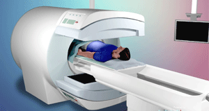
You’ll lie still on a table while an arm like device supporting a tracer-sensitive camera passes back and forth over your body. The scan itself can take up to an hour. The procedure is painless.
Orthopedic Specialist at Shreya Hospital in Ghaziabad might order a three-phase bone scan, which includes a series of images taken at different times. A number of images are taken as the tracer is injected, then shortly after the injection, and again 3 to 5 hours after the injection.
To better see some bones in your body, your doctor might order additional imaging called single-photon emission computerized tomography (SPECT). This imaging can help with conditions that are especially deep in your bone or in places that are difficult to see. During a SPECT scan, the camera takes images as it rotates around your body.
 Dr. Mishra’s Shahdara Orthopaedic Center Shahdara Orthopaedic Center, Best Orthopaedic Doctor in Shahdara, Delhi
